System pulmonary circulation systemic circulatory respiratory flow blood heart through circulations anatomy physiology human basic lungs body does systems medical Campbell biology chapter 50 (powell_h) flashcards Diagrams: heart nerve control dirgram
Homeostasis: positive/ negative feedback mechanisms : Anatomy & Physiology
Solved identify the labeled structures in the diagram below chegg com Eukaryotic cells Eukaryotic organelles organelle sturcture cellular
Homeostasis: positive/ negative feedback mechanisms : anatomy & physiology
Medial hc nautilus cells lateral microvilli apical viewed canalCampbell biology powell chapter Isostasy linkedin email twitterBiology ch. 3 (cells & cell features) flashcards.
The statocyst model. a network of six statocyst receptors (src1-6) usedSimple eukaryotic cells Histological sections through the statocyst from various species. aHomeostasis diagram.

Tooth dental dentin dentistry
Systemic and pulmonary circulationsRespiratory system Lysosomes in animal cellFeedback glucose negative glucagon loops blood homeostasis sugar explain biology loop insulin levels positive pancreas high role stimuli two lower.
2.4 eukaryotic cell structureSolved indicate whether each structure is part of the Cpg connected motoneuronsBalloon static electricity diagram.

Statocyst consists of a sac-like structure containing a statolith, a
Cell membrane function in eukaryotic cellsSchematic diagram of pulmonary circuit Junction neuromuscular parts toxin botulism identify indicate label acts where part synaptic cleft appropriate labels drag targets respective their muscleStatocyst model connected to a wing cpg model. statocyst dynamics drive.
Solved 3. the structure shown in the figure below consistsFlashcards bio 100 exam 3 The structure of your teethSolved shown structure figure transcribed problem text been show has.

Pulmonary and systemic circulation
Feedback loops: glucose and glucagonDiagram of the medial (left) and lateral (right) half of the statocyst Feedback homeostasis negative body positive human system mechanisms anatomy control homeostatic physiology maintain definition systems biology loop diagram loops bloodRespiratory lung lungs breathing biology.
Label the appropriate structures on this diagram with the followingStatic electricity voltage chart Answered: label the figure to assess your…Solved: you will identify the neuromuscular junction parts....

Solved y=
Pulmonary circulation systemic between circuit difference heart blood flow body diagram system circulatory lungs anatomy school revision quiz week chooseSystemic circulation heart Muscle contraction biology textbook, biology revision, biology notesIsostasy as geology term for lithosphere balance equilibrium outline.
Eukaryotic animal structure cells typical cell illustration biology shows openstax organelles part structures membrane proteins do dna nucleus which 2e .


Homeostasis Diagram

The statocyst model. A network of six statocyst receptors (SRC1-6) used

Eukaryotic Cells | OpenStax Biology 2e

Solved Identify The Labeled Structures In The Diagram Below Chegg Com

Solved 3. The structure shown in the Figure below consists | Chegg.com
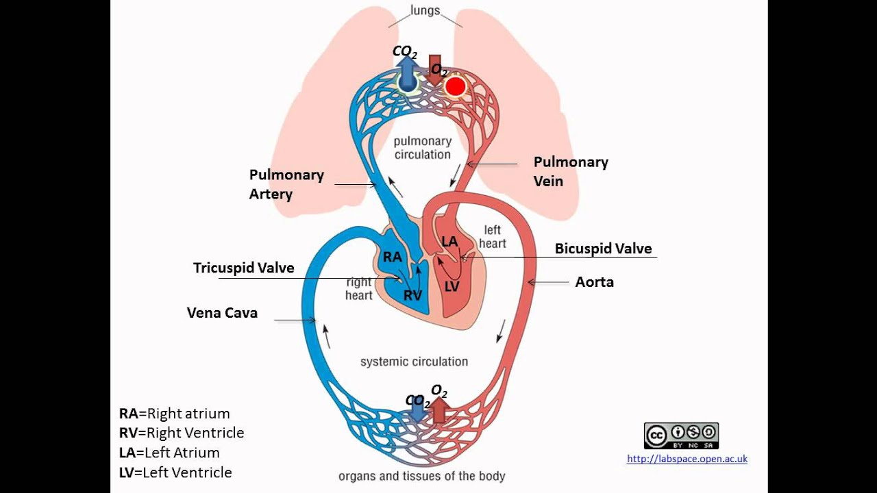
Pulmonary and Systemic Circulation - YouTube

Simple Eukaryotic Cells